Reconstructive and Functional Surgery
The field of Oculoplastic Surgery is based upon the knowledge and principles of Ophthalmology – to know, understand, and protect the eye. All eyelid surgery has the goal of safety, maintaining vision and comfort for the eye, with the ideal of maintaining the appearance of symmetry and aesthetics. The climate of Western Canada creates special demands in achieving this balance.
The dynamic nature of the eyelid means many abnormalities of eyelid position and function can occur. The eyelid may turn in (entropion), out (ectropion), droop (ptosis), or retract (lid retraction). Its exposure to sun can produce malignancies (basal cell carcinoma, squamous cell carcinoma, and melanoma). The environment of Western Canada means damage to many eyelids. The natural aging process can contribute to many eyelid changes, and the eyelids complicated development in the early months of pregnancy can produce various birth defects.
Tragically, eyes are still lost to trauma, tumors, and severe ocular diseases. The role of enucleation (removal of an eye) and the reconstructive work of socket repair using implants (e.g. Biomatrix, hydroxyapatite) are integral parts of Dr. Chan’s practice.
Orbital Disease and Thyroid Orbitopathy
An enormous spectrum of problems occur in the tissues around and behind the eye – the orbit.
The most common disease processes in the orbit are inflammatory diseases– Thyroid Orbitopathy and the idiopathic inflammatory “pseudotumors”. These are non-malignant processes that have an enormous range of presentations – acute or chronic, painless or miserable, easily responding to treatment or stubborn in their response. Many different treatments can be required – both surgical and medical therapies can be needed. Drugs such as corticosteroids (e.g. Cortisone or Prednisone) and other immunosuppressive and chemotherapeutic agents like methotrexate all have a role. Occasionally, Radiotherapy is used even though these are benign (non-cancerous) conditions. Drs. Chan and Johnson have long-established working relationships with specialists in these fields at the Cross Cancer Clinic and other hospitals in Edmonton and Alberta to allow this team approach.
Many other types of problems also occur in the orbit – tumors both benign and malignant, congenital abnormalities (birth defects), and diseases which spread from the adjacent sinuses and brain.
Clinical experience is the most important part of the assessment of these problems and their management techniques. The excellent CT scan and MRI techniques specific for the orbit allow many diagnoses without intervention. Also, the need for precise diagnosis may require tissue analysis and Drs. Chan and Johnson have developed teamwork with a dedicated ophthalmological pathologist for optimal diagnosis.
Thyroid Eye Disease
Thyroid eye disease has many names – thyroid associated orbitopathy, Grave’s disease, Grave’s orbitopathy, etc. All these terms mean the same thing – inflammation of the tissues around and behind the eye producing varying amounts of swelling and scarring.
Thyroid eye disease is a descriptive term for swelling of the tissues around and behind the eye. It is an autoimmune disease (i.e.: where your body attacks itself) and there are at least various theories to explain its cause and development. Thyroid eye disease usually, but not always, occurs in people with a history of thyroid problems. However, thyroid eye disease can occur decades before, or decades after, the development of thyroid gland disease. Thyroid eye disease has not been consistently shown to be caused by radioactive iodine or thyroid surgery but there is concern that radioactive iodine can be a negative influence in some people. A great deal of research is being conducted all over the world trying to obtain more information on the cause of thyroid eye disease. Interestingly, smoking definitely makes thyroid eye disease worse.
Thyroid eye disease has many effects. Most people, in the development of thyroid eye disease, will relate that their eyes are sore, gritty, burning and irritable. Generally, there is swelling of the eyelids and the tissues around the eye and some people develop double vision (diplopia). The swelling of the tissues is caused by inflammation of the eye muscles behind the eye, with some inflammation of the fat behind the eye (the eye sits in a “shock absorber” of special orbital fat). The inflammation causes swelling of the muscles and the fat and this causes the appearance of proptosis – bulging forward of the eyes – and also can cause symptoms of double vision. Occasionally, the swelling of the muscles is so severe that they squeeze the optic nerve (the vision nerve) deep behind the eye and actually “strangle” the nerve and produce loss of vision. Fortunately, this very serious problem usually responds to treatment
The general management of thyroid eye disease is based on the experience that usually the eye disease is active for 12 – 18 months and then naturally stabilizes. Maximum comfort can be obtained by the use of drops and ointment. The eyes are usually irritable and sore, partly because they are proptotic / bulging / exposed. Eye drops during the day will keep the eyes more comfortable. Also, patients frequently sleep with their eyes open and need ointment to the eyes at bedtime so the eyes do not dry out overnight. A bedroom humidifier can also be useful.
Thyroid eye disease tends to evolve over a time period of six – twelve – eighteen months and this is a very individualized process. Then, the disease process tends to stop (burn itself out) and the changes stabilize and occasionally almost return to normal.
It can be possible to modify or reduce the progression of thyroid eye disease. The standard treatment for active, severe thyroid eye disease is high dose cortisone – this usually works but has a multitude of possible side effects (e.g. diabetes, osteoporosis with broken bones, weight gain, muscle loss, acne, cataracts, ulcers, intestinal bleeding etc.). This therapy is usually helpful but with its significant potential for complications is used only when absolutely necessary. Another traditional way to stop the inflammation of the thyroid eye disease is with radiation to the orbits. This can be an effective way of settling down the inflammation and bringing the disease process under control, using only about one third the dose that cancer patients require. But there still can be potential complications, and controversy about its effectiveness persists.
There are a variety of different treatments which have been recommended in various places in the world over the years, but the consensus is that high dose cortisone is the most effective and relatively safest treatment for the severe patient. However controversies remain. Drugs that modify the immune system – like methotrexate – also show promise in helping to control the disease with minimum side effects. (Methotrexate is very commonly used for other immune diseases like rheumatoid arthritis).
Once the disease has stabilized, and occasionally if there are severe problems before the disease has stabilized, surgical management of the tissues around the eye can be considered.Consequently, most patients are best served by waiting out the evolution and changes of the thyroid eye disease. During this time, eye drops and ointment are the mainstay of treatment. Also, some patients will require humidifiers at home or in the workplace to improve humidity in the atmosphere, and occasionally patients will benefit by wearing glasses or goggles to protect their corneas.
Orbital decompression is a technique used to remove the bones of the orbit behind the eye to relieve pressure on the tissues behind the eye. This can be performed for the preservation of vision, but also is used to restore the patient’s comfort and appearance by reducing the bulging (proptosis) of the eye. For the rare patient who is losing vision because of compression of the optic nerve, orbital decompression is sight saving. Also, this can be done for patients who are very proptotic (very bulging eyes). The operation usually takes about 90 minutes for both eyes and usually requires 18 – 24 hours in the hospital. This is a successful and relatively safe operation, but it is well known that approximately 1/4 of patients will have development of double vision (or more commonly worsening of their previously existing double vision).
To straighten the eyes and treat the double vision, strabismus surgery is required. The double vision in thyroid eye disease patients is caused by swelling of the eye muscles which then causes scarring with contracture of muscles which pull the eyes into an abnormal position producing double vision, or by limiting the normal eye movements so that there is double vision when the eyes are in different directions of gaze i.e. when the eyes move around. Most commonly this occurs with the eyes in the position of upgaze, and outward (lateral) gaze. One has to realize that double vision, although a nuisance, is not truly important to one’s existence as long as the straight ahead position and downwards reading position do not have double vision. If the double vision is only in far upgaze or lateral (side) gaze, then the general advice is to simply live with this.
Initially, before the disease has stabilized, the double vision is treated by simply occluding or covering one eye. Unfortunately, recent laws have stated that even though there is no double vision with one eye covered, a patient cannot drive a vehicle until he/she has had one eye covered for 3 months.There is no point in straightening the eye if the thyroid eye disease has not stabilized, for this could require yet another operation later on. Consequently, the double vision, although very unsatisfactory, has to be tolerated until the thyroid eye disease has been stabilized. Then, the eyes can almost invariably be brought back into alignment with strabismus surgery. Generally, the strabismus surgery is done with the patient asleep, and often, the adjustable suture technique is used – whereby, the operation is done as planned with the patient asleep and then after the patient has recovered, the fine tuning on the eye muscle is performed. This technique is very useful because of the marked unpredictability of the thyroid eye muscles.
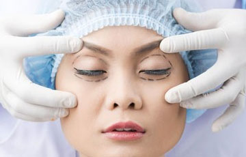
The third step in the management of thyroid eye disease is correction of the eyelids. The upper lid may need to be dropped, and the lower lid may need to be raised. This is not done just for appearance of the eyes – but also to maximize comfort and improve the protection to the eyes. Generally, these manipulations of the eyelids are done under local anesthetic and with an anesthetist present to make the procedure comfortable. Like straightening the eye muscles to correct double vision the thyroid eyelids are notorious in their unpredictability. However, using a variety of different techniques almost always the eyelids can be repositioned to achieve a satisfactory lid position, and consequently, normal eye comfort and safety.
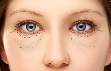
The final step for some thyroid eye disease patients is to remove any extra “baggy eyelids” which have been present. Similar to the setting of the eyelid height, this is usually done under local anesthetic, with appropriate sedation by an anesthetist. This is a cosmetic procedure.
In summary, thyroid eye disease is a challenging and not very well understood problem. However, by following the principles of allowing the thyroid eye disease to evolve and then correcting whatever problems are present, the vast majority of patients can be safely and effectively brought through the thyroid eye disease. A minority of patients will require urgent management to preserve their vision. Every patient with thyroid eye disease is an individual – the variation in thyroid eye disease is truly amazing.
Lacrimal Disease (Tearing)
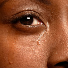
The lacrimal system is the tear duct system – to drain the normal tears away from the eye. There is a need for constant balance- too many tears produce the annoying symptoms of blurred vision and a wet face – too few tears create the misery of dry eyes.
There are two general causes of tearing – (1) abnormalities of the tear drainage system (Nasolacrimal Duct – NLD) and (2) reflex tearing whereby any irritation to the eyes causes reflex tear production (i.e. wind, dust, pollution). Dr. Johnson has special expertise in tear duct surgery, but before the tear outflow system can be surgically corrected it is critical to ensure that the cause is poor drainage (outflow obstruction). The most common cause of annoying reflex tearing is the dry eye syndrome!! and of course surgery to improve the tear drainage system will worsen an existing dry eye. Drs. Chan and Johnson can generally solve a drainage problem, but there is no cure for dry eye, only some symptom improvement.
Lacrimal surgery is frequently required on infants – the common Congenital Nasolacrimal Duct Obstruction of infants (please contact our office for a detailed explanatory pamphlet).
Also lacrimal surgery can be required at virtually any age of life – the tear ducts can become blocked from many reasons – trauma, radiation, other types of operations (e.g. sinus surgery) and the most common cause – unknown!
Evaluation of the tear duct is based upon history, and also the anatomy of the tear duct. This is assessed by the simple NLD irrigation performed in the office using just eye drops for freezing. This is not a treatment – it just allows the Doctor to identify the presence and location of a blockage.
Tear duct surgery generally tries to rebuild the natural tear duct into the nose – i.e. DCR (dacryocystorhinostomy). Frequently (depending on the location of the blockage) temporary tear duct tubes are placed as part of the DCR – these are not drainage tubes, but rather are ‘stents’ used to keep the tear duct stretched open against the scar tissue which occurs during healing. This operation can be done either through the skin (leaving a small mark) or through the nose i.e. endonasal DCR – with no skin incision but perhaps a lower success rate.
Sometimes it is not possible to use the natural tissues to rebuild the tear duct and then a permanent artificial tear duct is created using the pyrex glass Lester Jones tube – this is the conjunctival DCR (CDCR). These Jones tubes are usually successful, and are much better than chronic tearing, but like any artificial system, maintenance can be required.
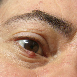
Eyelid Lesions – Lumps and Bumps
There are an astonishing number of tumors that can affect the tissues around the eye (the eyelids and orbit), and inside the eye itself. While Drs. Chan and Johnson share the investigation of tumors that develop inside the eye with other colleagues, they are frequently called upon for help with surgical management.
Dr. Chan’s and Dr. Johnson’s expertise in oculoplastic and orbital surgery means that a great variety of tumors around and behind the eye – both benign and malignant – are referred for management.
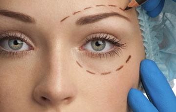
There are multiple benign lid tumors and these usually require straightforward surgical treatment and are commonly of only cosmetic significance. The commonly seen skin cancers (basal cell carcinoma, squamous cell carcinoma) are generally best treated with eyelid surgery and reconstruction, and occasionally more complicated tumors require Drs. Chan and Johnson to work in teamwork with the specialists at the Cross Cancer Clinic.
Orbital tumors are part of this area of interest and expertise. So many possibilities and presentations exist in this field that only individual examinations and consultations are appropriate.